NEW ASSOCIATE MEMBERSHIP FOR DEVELOPING COUNTRIES

Reduced fees for SCMR membership - click here for more information.


SCMR-LAC Multicenter Registry is enrolling!

Contact us at jlaraf@fcm.unicamp.br to participate - Download the Instructional Manual here

SCMR-LAC on Twitter - follow us: http://twitter.com/scmrlac


Nov 29, 2009

MVO as an endpoint


A couple of posts before we mentioned how CMR would become an important endpoint in clinical studies. This is another classical point in this regard. As soon as more sites prove their consistency in research, your cases/month can go up significantly. Participating in research always adds to your institution and generates more income.

J Am Coll Cardiol. 2009 Dec 1;54(23):2145-2153.

Impact of Primary Coronary Angioplasty Delay on Myocardial Salvage, Infarct Size, and Microvascular Damage in Patients With ST-Segment Elevation Myocardial Infarction Insight From Cardiovascular Magnetic Resonance.

Francone M, Bucciarelli-Ducci C, Carbone I, Canali E, Scardala R, Calabrese FA, Sardella G, Mancone M, Catalano C, Fedele F, Passariello R, Bogaert J, Agati L.

Cardiovascular Magnetic Resonance Unit, Department of Radiology Sciences, "Sapienza" University of Rome, Rome, Italy.

OBJECTIVES: We investigated the extent and nature of myocardial damage by using cardiovascular magnetic resonance (CMR) in relation to different time-to-reperfusion intervals. BACKGROUND: Previous studies evaluating the influence of time to reperfusion on infarct size (IS) and myocardial salvage in patients with ST-segment elevation myocardial infarction (STEMI) have yielded conflicting results. METHODS: Seventy patients with STEMI successfully treated with primary percutaneous coronary intervention within 12 h from symptom onset underwent CMR 3 +/- 2 days after hospital admission. Patients were subcategorized into 4 time-to-reperfusion (symptom onset to balloon) quartiles: 90 to 150 min (group II, n = 17), >150 to 360 min (group III, n = 17), and >360 min (group IV, n = 17). T2-weighted short tau inversion recovery and late gadolinium enhancement CMR were used to characterize reversible and irreversible myocardial injury (area at risk and IS, respectively); salvaged myocardium was defined as the normalized difference between extent of T2-weighted short tau inversion recovery and late gadolinium enhancement. RESULTS: Shorter time-to-reperfusion (group I) was associated with smaller IS and microvascular obstruction and larger salvaged myocardium. Mean IS progressively increased overtime: 8% (group I), 11.7% (group II), 12.7% (group III), and 17.9% (group IV), p = 0.017; similarly, MVO was larger in patients reperfused later (0.5%, 1.5%, 3.7%, and 6.6%, respectively, p = 0.047). Accordingly, salvaged myocardium markedly decreased when reperfusion occurred >90 min of coronary occlusion (8.5%, 3.2%, 2.4%, and 2.1%, respectively, p = 0.004). CONCLUSIONS: In patients with STEMI treated with primary percutaneous coronary intervention, time to reperfusion determines the extent of reversible and irreversible myocardial injury assessed by CMR. In particular, salvaged myocardium is markedly reduced when reperfusion occurs >90 min of coronary occlusion.

Too little, too far


The results of the poll shows that valvular heart disease assessment by CMR is still very much underused in Latin America. We (and I mean the worldwide CMR community) still have a long way to go...

Nov 23, 2009

Delayed enhancement in CMR: the "gold standard" for prognosis?


A couple of years ago many people debated whether CMR was the gold standard for viabiliby. Then came the gold standard for detection of infarcts. Are we into a new era as DLE stands for the gold standard of prognosis? In very wide and different scenarios DLE always seems to add prognostic information. This article by Dr. Cheong et al adds another point on the graph:

http://circ.ahajournals.org/cgi/content/abstract/120/21/2069?etoc


Prognostic Significance of Delayed-Enhancement Magnetic Resonance Imaging
Survival of 857 Patients With and Without Left Ventricular Dysfunction
Benjamin Y.C. Cheong, MD; Raja Muthupillai, PhD; James M. Wilson, MD; Angela Sung; Steffen Huber, MD; Samir Amin, BA; MacArthur A. Elayda, MD, PhD; Vei-Vei Lee, MS; Scott D. Flamm, MD

From the Departments of Radiology (B.Y.C.C., R.M., S.H., A.S., S.D.F.), Cardiology (B.Y.C.C., J.M.W., S.D.F.), and Biostatistics and Epidemiology (M.A.E., V.V.L.), the Texas Heart Institute at St. Luke’s Episcopal Hospital, and the Departments of Medicine (B.Y.C.C., S.A.) and Radiology (B.Y.C.C., R.M., S.D.F.), Baylor College of Medicine, Houston, Tex. Dr Flamm is currently at the Cleveland Clinic Foundation, Cleveland, Ohio.

Correspondence to B.Y.C. Cheong, MD, Department of Diagnostic and Interventional Radiology, St. Luke’s Episcopal Hospital and the Texas Heart Institute, 6720 Bertner Ave, MC 2–270, Houston, TX 77030. E-mail bcheong@sleh.com

Received January 20, 2009; accepted September 11, 2009.

Background— Left ventricular ejection fraction is a powerful independent predictor of survival in cardiac patients, especially those with coronary artery disease. Delayed-enhancement magnetic resonance imaging (DE-MRI) can accurately identify irreversible myocardial injury with high spatial and contrast resolution. To date, relatively limited data are available on the prognostic value of DE-MRI, so we sought to determine whether DE-MRI findings independently predict survival.

Methods and Results— The medical records of 857 consecutive patients who had complete cine and DE-MRI evaluation at a tertiary care center were reviewed regardless of whether the patients had coronary artery disease. The presence and extent of myocardial scar were evaluated qualitatively by a single experienced observer. The primary, composite end point was all-cause mortality or cardiac transplantation. Survival data were obtained from the Social Security Death Index. The median follow-up was 4.4 years; 252 patients (29%) reached one of the end points. Independent predictors of mortality or transplantation included congestive heart failure, ejection fraction, and age (P<0.0001 for each), as well as scar index (hazard ratio, 1.26; 95% confidence interval, 1.02 to 1.55; P=0.033). Similarly, in subsets of patients with or without coronary artery disease, scar index also independently predicted mortality or transplantation (hazard ratio, 1.33; 95% confidence interval, 1.05 to 1.68; P=0.018; and hazard ratio, 5.65; 95% confidence interval, 1.74 to 18.3; P=0.004, respectively). Cox regression analysis showed worse outcome in patients with any DE in addition to depressed left ventricular ejection fraction (<50%).

Conclusion— The degree of DE detected by DE-MRI appears to strongly predict all-cause mortality or cardiac transplantation after adjustment for traditional, well-known prognosticators.

Nov 22, 2009

Dyssynchrony analysis - a real niche for CMR


Dyssynchrony analysis is a new niche were CMR can provide a very comprehensive contribution by adding not only quantitative information but DLE visualization as well. The integration of data in this regard is a unique feature of CMR over all other methods. Paper pointed out by Dr. Florangel Martínez

Radiology. 2009 Nov;253(2):364-71. Epub 2009 Jul 31.
Interventricular mechanical dyssynchrony: quantification with velocity-encoded MR imaging.


Muellerleile K, Baholli L, Groth M, Barmeyer AA, Koopmann K, Ventura R, Koester R, Adam G, Willems S, Lund GK.

Center for Cardiology and Cardiovascular Surgery, University Medical Center Hamburg-Eppendorf, Martinistrasse 52, D-20246 Hamburg, Germany. ka.muellerleile@uke.de

PURPOSE: To evaluate the performance of velocity-encoded (VENC) magnetic resonance (MR) imaging, as compared with pulsed-wave echocardiography (PW-ECHO), in the quantification of interventricular mechanical dyssynchrony (IVMD) as a predictor of response to cardiac resynchronization therapy (CRT). MATERIALS AND METHODS: The study was approved by the local ethics committee, and all patients provided written informed consent. The study involved the examination of 45 patients (nine women, 36 men; median age, 60 years; interquartile age range, 47-69 years) with New York Heart Association class 2.0-3.0 heart failure and a reduced left ventricular ejection fraction (median, 25%; interquartile range, 21%-32%), with (n = 25) or without (n = 20) left bundle branch block. Aortic and pulmonary flow curves were constructed by using VENC MR imaging and PW-ECHO. IVMD was defined as the difference between the onset of aortic flow and the onset of pulmonary flow. Intraclass correlation coefficient, Spearman correlation coefficient, Bland-Altman, and Cohen kappa analyses were used to assess agreement between observers and methods. RESULTS: Inter- and intraobserver agreement regarding VENC MR imaging IVMD measurements was very good (intraclass r = 0.96, P < .001; mean bias, -3 msec +/- 11 [standard deviation] and 0 msec +/- 10, respectively). A strong correlation (Spearman r = 0.92, P < .001) and strong agreement (mean difference, -6 msec +/- 16) were found between VENC MR imaging and PW-ECHO in the quantification of IVMD. Agreement between VENC MR imaging and PW-ECHO in the identification of potential responders to CRT was excellent (Cohen kappa = 0.94). CONCLUSION: VENC MR measurements of IVMD are equivalent to PW-ECHO measurements and can be used to identify potential responders to CRT. (c) RSNA, 2009.

Nov 15, 2009

Cases Needed!


Submit your case to the Medis/SCMR SAT - DEADLINE Dec 1.

Need to be a SCMR member!


Juliano

>>>
Call for Cases: Medis/SCMR Self-Assessment Test 2010

Dear SCMR Member,

A popular and effective means of education, the Medis/SCMR Self-Assessment Test will be featured at the SCMR 2010 for the tenth consecutive year.
We are calling on you to share your interesting cardiac MR cases with other SCMR members through the Medis/SCMR Self-Assessment Test, so they too can learn from them.
The cases you provide will be peer-reviewed and you will be notified by e-mail if they are accepted. Cases used in the test at the SCMR 2010 will mention your name and affiliation.
Fast Online Submission of Cases
The online submission process consists of just five steps, allowing you to submit your cases in a few minutes' time.
1. Access the online Self-Assessment Center at http://scmr.medis.tv/
You can submit your cases using MS Internet Explorer version 6, 7 or 8.
2. If you are a first-time visitor, create your account and continue with submitting cases right away—no need to wait for your password.
3. Click 'Add new case' and provide a case description, a test question and three to six possible answers.
4. Add one to four images (JPG) or movies (AVI or MPG).
5. Preview and save your case.
You can add up to four cases. After you have submitted your cases, you can return to the online Self-Assessment Center any time to edit your cases, remove cases or to check their review status. You can access online help by clicking 'Manual' in the top right corner of the page.
Submission Deadline and More Info
Please submit your cases before December 1, to allow enough time for case review. If you have any questions or comments, please send an e-mail to info@medis.nl.

CMR Endpoints in Trials


Due to the accuracy and reduced variability, we should be seeing more and more in the use of CMR variables as endpoints in different trials. Of course the myocardium will always be a target but one cannot forget vascular wall assessment or functional data as well.

J Am Coll Cardiol. 2009 Nov 3;54(19):1787-94.
Effects of high-dose modified-release nicotinic acid on atherosclerosis and vascular function: a randomized, placebo-controlled, magnetic resonance imaging study.

Lee JM, Robson MD, Yu LM, Shirodaria CC, Cunnington C, Kylintireas I, Digby JE, Bannister T, Handa A, Wiesmann F, Durrington PN, Channon KM, Neubauer S, Choudhury RP.

Department of Cardiovascular Medicine, University of Oxford and Oxford Centre for Clinical Magnetic Resonance Research (OCMR), Oxford, United Kingdom.

Comment on:

* J Am Coll Cardiol. 2009 Nov 3;54(19):1795-6.

OBJECTIVES: Our aim was to determine the effects of high-dose (2 g) nicotinic acid (NA) on progression of atherosclerosis and measures of vascular function. BACKGROUND: NA raises high-density lipoprotein cholesterol (HDL-C) and reduces low-density lipoprotein cholesterol and is widely used as an adjunct to statin therapy in patients with coronary artery disease. Although changes in plasma lipoproteins suggest potential benefit, there is limited evidence of the effects of NA on disease progression when added to contemporary statin treatment. METHODS: We performed a double-blind, randomized, placebo-controlled study of 2 g daily modified-release NA added to statin therapy in 71 patients with low HDL-C (<40 mg/dl) and either: 1) type 2 diabetes with coronary heart disease; or 2) carotid/peripheral atherosclerosis. The primary end point was the change in carotid artery wall area, quantified by magnetic resonance imaging, after 1 year. RESULTS: NA increased HDL-C by 23% and decreased low-density lipoprotein cholesterol by 19%. At 12 months, NA significantly reduced carotid wall area compared with placebo (adjusted treatment difference: -1.64 mm(2) [95% confidence interval: -3.12 to -0.16]; p = 0.03). Mean change in carotid wall area was -1.1 +/- 2.6 mm(2) for NA versus +1.2 +/- 3.0 mm(2) for placebo. In both the treatment and placebo groups, larger plaques were more prone to changes in size (r = 0.4, p = 0.04 for placebo, and r = -0.5, p = 0.02 for NA). CONCLUSIONS: In statin-treated patients with low HDL-C, high-dose modified-release NA, compared with placebo, significantly reduces carotid atherosclerosis within 12 months. (Oxford Niaspan Study: Effects of Niaspan on Atherosclerosis and Endothelial Function; NCT00232531).

CMR conferences in Brazil


In 2010 we will have 6 big opportunities to meet and discuss CMR: 3 in the first semester and 3 in the second (dates to be announced soon). Interestingly, half organized by radiology and half by cardiologists, a definite plus in keeping both groups integrated. The focus of each meeting should be a little bit different, taking into account the public, the main objectives and the form of the meetings. Nevertheless, we believe the country has been very active and will continue to do so in the foreseeable future.

Nov 5, 2009

CMR and Angios go together


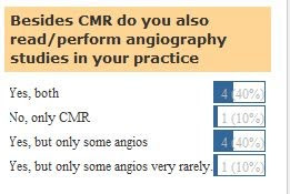
As a result of our poll, most CMR physicians perform both heart and angio studies. This is a very important issue since training in CMR has to take this point in consideration and make sure that fellows get enough exposure to the analysis of central and peripheral angio studies, enhancing their role as cardiovascular CMR imagers.

Nov 2, 2009

SCMR-LAC 2009 Annual Meeting


With the exceptional hospitality of Dr. Erick Alexanderson and all Mexican cardiologists, the SCMR-LAC 2009 Annual Meeting took place in Puebla, Nov 31. The meeting welcomed the presence of specialists from other areas with a very rich discussion on improvements in the chapter. Dr. Meave and Dr. Morelos as well as Dr. De LaPena inputs were very positive and we should provide a summary of the discussions very briefly in the site.

In the name of the chapter we openly thank the Mexican Society of Cardiology and Dr. Alexanderson for such a wonderful opportunity.