NEW ASSOCIATE MEMBERSHIP FOR DEVELOPING COUNTRIES

Reduced fees for SCMR membership - click here for more information.


SCMR-LAC Multicenter Registry is enrolling!

Contact us at jlaraf@fcm.unicamp.br to participate - Download the Instructional Manual here

SCMR-LAC on Twitter - follow us: http://twitter.com/scmrlac


Dec 29, 2010

Happy New Year


This is the last post for 2010. All we have to say is that we wish all readers a very happy and productive New Year!

Nothing beats hard work and we will be driven by that motto all year around in 2011.

Best wishes, Juliano.

Dec 28, 2010

3D black blood coronary imaging wiht near 100% tracking


If widely reproducible and applicable in a clinical routine, this could mean a significant change in the way coronary angios are done with CMR by significantly shortening the acquisition time. In real life, if you get 20-30% respiratory efficiency you are a lucky physician.

Link here.


High-resolution 3D coronary vessel wall imaging with near 100% respiratory efficiency using epicardial fat tracking: Reproducibility and comparison with standard methods.

Scott AD, Keegan J, Firmin DN.

Cardiovascular Magnetic Resonance Unit, National Heart and Lung Institute, Imperial College London, London, United Kingdom; Cardiovascular Magnetic Resonance Unit, Royal Brompton and Harefield NHS Foundation Trust, London, United Kingdom.

J. Magn. Reson. Imaging 2011;33:77-86

Dec 4, 2010

It's not rocket science you know - part I


A must read for all involved in the field. This was commented on the scmr.org news section as well - I try not to repeat manuscripts but this one is really key if you are beginning in CMR or want to explain to your clinicians how spins will generate images.

Free full text at: http://jcmr-online.com/content/12/1/71

Nov 27, 2010

LGE in papillary muscle in myocardial infarction


An astonishing 40% of patients investigated in this Japanese study after an STEMI had signs of LGE in the papillary muscles. The clinical significance however was not so important since this finding was not associated with mitral regurgitation or ventricular remodeling.

Read the abstract here.

Nov 21, 2010

Valve disease : CMR as the reference method

On the other hand (see previous post), a recent manuscript in Circ Cardiovasc Imaging on mitral regurgitation has compared 2D and 3D TE echo with the reference method - yes, CMR...

If only we could convince ourselves!

Circ Cardiovasc Imaging. 2010 Nov 1;3(6):694-700. Epub 2010 Sep 1.
Quantitative Assessment of Mitral Regurgitation: Comparison Between Three-Dimensional Transesophageal Echocardiography and Magnetic Resonance Imaging.


Shanks M, Siebelink HM, Delgado V, van de Veire NR, Ng AC, Sieders A, Schuijf JD, Lamb HJ, Ajmone Marsan N, Westenberg JJ, Kroft LJ, de Roos A, Bax JJ.

Department of Cardiology and the Department of Radiology, Leiden University Medical Center, Leiden, The Netherlands; and the Department of Cardiology Rijnland Ziekenhuis, Leiderdorp, The Netherlands.
Abstract

Background- Quantification of mitral regurgitation severity with 2-dimensional (2D) imaging techniques remains challenging. The present study compared the accuracy of 2D transesophageal echocardiography (TEE) and 3-dimensional (3D) TEE for quantification of mitral regurgitation, using MRI as the reference method. Methods and Results- Two-dimensional and 3D TEE and cardiac MRI were performed in 30 patients with mitral regurgitation. Mitral effective regurgitant orifice area (EROA) and regurgitant volume (Rvol) were estimated with 2D and 3D TEE. With 3D TEE, EROA was calculated using planimetry of the color Doppler flow from en face views and Rvol was derived by multiplying the EROA by the velocity time integral of the regurgitant jet. Finally, using MRI, mitral Rvol was quantified by subtracting the aortic flow volume from left ventricular stroke volume. Compared with 3D TEE, 2D TEE underestimated the EROA by a mean of 0.13 cm(2). In addition, 2D TEE underestimated the Rvol by 21.6% when compared with 3D TEE and by 21.3% when compared with MRI. In contrast, 3D TEE underestimated the Rvol by only 1.2% when compared with MRI. Finally, one third of the patients in grade 1 and ≥50% of the patients in grade 2 and 3, as assessed with 2D TEE, would have been upgraded to a more severe grade, based on the 3D TEE and MRI measurements. Conclusions- Quantification of mitral EROA and Rvol with 3D TEE is feasible and accurate as compared with MRI and results in less underestimation of the Rvol as compared with 2D TEE.

Underuse of CMR for valve assessment


Despite the high prevalence of valve disease in Latin America, CMR is still underused according to our responders.

Nov 8, 2010

Quantifying LGE in HCM


What should be the threshold for LGE quantification in HCM? According to Harrigan > 6SD is the most reproducible and closest to visual assessment than other numbers.

Semiautomation in these cases should really help to establish a number in which to follow patients longitudinally.

Read more at: http://radiology.rsna.org/content/early/2010/10/28/radiol.10090526.long

Nov 2, 2010

Move over 3.0T...


Just as we were warming up, 7.0T comes to town.

An interesting manuscript on the use of this new high field imaging in coronary arteries.

Link here.

Oct 25, 2010

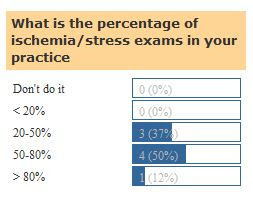
Ischemia still leads the way


According to our visitors, a great portion of the exams in their sites are ischemic testing.

Oct 23, 2010

Valvular heart disease: under used by CMR?


This review mentions many applications of CMR in assessing cardiac valve disease. This is a field where CMR is clearly underused and should gain further terrain as knowledge advances.

Reach the link here.

Oct 18, 2010

Pulmonary Hypertension by CMR


The use of CMR to assess the right ventricle is undisputed. However, there is much more to the RV in pulmonary hypertension. This manuscript reviews all that can be achieved in PH with CMR. Widely recommended.

Link here.

Oct 13, 2010

ECG prediction, PCI and CMR in acute myocardial infarction


A very interesting manuscript in Spanish from the Rev Esp Cardiol with an editorial by Dr Raymond Kwong and Tomas Neilan.

The full PDF is here.

La suma de la elevación del segmento ST predice mejor la obstrucción microvascular en pacientes tratados con éxito con una intervención coronaria percutánea primaria. Un estudio de resonancia magnética cardiovascular
Oliver Husser. Vicente Bodí. Juan Sanchis. Julio Núnez. Luis Mainar. Eva Rumiz. María Pilar López-Lereu. José Monmeneu. Fabián Chaustre. Isabel Trapero. María J. Forteza. Günter A.J. Riegger. Francisco Javier Chorro. Àngel Llàcer.
Rev Esp Cardiol.2010; 63 :1145-54

Oct 6, 2010

Stress Perfusion Review


A review by Christiansen et al in Heart, Lung and Circulation provides a thorough look into the clinical and technological aspects of CMR perfusion.

Link here.

Sep 19, 2010

3 Important reviews


This week 3 important reviews were published:

1. Iron Overload Cardiomyopathy Better Understanding of an Increasing Disorder.

Gujja P, Rosing DR, Tripodi DJ, Shizukuda Y. J Am Coll Cardiol. 2010 Sep 21;56(13):1001-1012.

2. Ischaemic heart disease assessment by cardiovascular magnetic resonance imaging.

Raj V, Agrawal SK. Postgrad Med J. 2010 Sep;86(1019):532-40.

3. Cardiovascular magnetic resonance evaluation of the patient with known or suspected coronary artery disease.

Schmid M, Daniel WG, Achenbach S. Heart. 2010 Oct;96(19):1586-92.

Follow the links to see the abstracts.

Sep 15, 2010

ANEURISMA DE PARED LATERAL UN HALLAZGO NO TAN FRECUENTE EN PACIENTES CHAGASICOS


Bertolasi y col, describen que el aneurisma ventricular apical izquierdo de la enfermedad de Chagas, que es característico y único de la miocardiopatía asociada con esta dolencia, presenta una incidencia del 60 al 65% en las diferentes series, vale decir una incidencia mayor que la del aneurisma de la cardiopatía isquemica(del 20 al 25%). En ambas entidades se observa la misma frecuencia de aparición de complicaciones relacionadas con la presencia del aneurisma ventricular: muerte súbita, arritmias ventriculares malignas y tromboembolismo pulmonar y sistémico.
La literatura revela que la localizacion más frecuente corresponde al ápice del ventrículo, si bien el aneurisma ha sido descrito en el septum interventricular y en las paredes posterior, inferior y apicoseptal de dicho ventrículo. Esta lesion ha sido hallada aislada en el 66,6% de los casos y asociada con un aneurisma en el ventrículo derecho en el 33,3%.
Mostramos un caso de un paciente chagasico, masculino de 70 años de edad, campesino, rescatado de muerte súbita y que posteriormente fue a implante de desfibrilador, que mostro a través de la RM un aneurisma de pared lateral, con fibrosis importante,revelando claramente el origen de las arritmias que desencadenaron el evento.
DRS JOAQUIN CANABAL, RAMON AGUILAR Y FLORANGEL MARTINEZ.
UNIDAD DE IMAGENES DEL CENTRO CLINICO VALENTINA CANABAL.BARQUISIMETO.VENEZUELA

Sep 13, 2010

New SCMR-LAC Newsletter


Just released.

Link here.

Fat in ARVD/C: let it go...


Most respondents do not regularly look for fat infiltration in ARVD/C. This is line with the recent guidelines in where CMR can aid in the diagnosis. It is a rather strange feeling to see that since the search for fat in ARVD/C was initially the most thought of use of CMR in the past. What a long way since then it has been.

Sep 5, 2010

CMR in Ischemic Cardiomyopathy Review


Just out in Heart by A M Beek et al. The authors describe not only the already known use of CMR for viability but new concepts on use of CMR for indications of ICD/CRT, grey zone and reconstructive surgery.

Heart. 2010 Sep;96(18):1494-501. Link here.

Aug 29, 2010

What's going on at the ESC 2010 in CMR


From the topics listed below, the focus has been on hands-on sessions and the use of CMR in heart failure and myocarditis. Personally, a bit disappointed to see so little of CMR represented in the meeting.

CLICK TO ENLARGE

Aug 19, 2010

While talking about T2*


Its use represents one of the most significant reductions in death in particular patient groups. Luckily, most of our readers are already using it regularly, despite some only more rarely.

T2* LatAm Meeting


In August 16/17 Drs. Dudley Pennell, Gillian Smith and Taigang He from the Royal Bromptom lead a course for over 25 Latin American physicians on the use of CMR for T2* imaging of the heart and liver in Campinas, Brazil. The results of this course will lead to major standardizations of acquisitions and reading of these exams helping to augment the availability of CMR to thalassemia patients in the region.

Aug 10, 2010

Dilated Cardiomyopathy and CMR


A thorough review in Circulation regarding dilated cardiomyopathies covers in depth the use of CMR in these conditions. Interestingly the review is focused on clinicians but diagnostic approaches are very much covered by CMR. A definite citation article where CMR is regarded as having a central role.

Link to the manuscript: http://circ.ahajournals.org/cgi/content/full/122/5/527

Jul 26, 2010

BioImage Study - role for CMR in primary prevention?


Part of the High-Risk Plaque (HRP) Initiative the BioImage study uses CMR to assess both atherosclerosis in the aorta and carotids. I also integrates CT, IMT, US and ABI markers and has already enrolled over 6800 patients.

The study is accepting new proposals from institutions to participate in the initiative. Further details can be found at:

http://www.hrpinitiative.com/hrpinit/bioimagerfp.jsp

Jul 20, 2010

Not unanimous but yet widely accepted


Most of the readers believe CMR should be performed in every patient with HCM. Based on the latest data published recently it is hard to argue against that.

Jul 12, 2010

CMR and aortic valve disease: new prognostic information


New prognostic information regarding CMR and severe aortic valve disease was published by a Brazilian group conducted by Dr. Clerio Azevedo. Should we only indicate aortic sugergy in patients with small amounts of fibrosis? A tough question that is raised by the paper and should generate more use of CMR in these conditions.

Azevedo CF et al. JACC 2010;56:278-87

Jul 5, 2010

Assessment of Myocardial Fibrosis Review


A paper in JACC commenting on how to measure myocardial fibrosis, a very important issue involving many cardiac diseases. CMR is represented by LGE, T1 mapping and tissue tagging.

Link here.

Jun 28, 2010

Cardiac Tumors


Most users see 1-10 tumors/year. That is a rather small number considering the incidence described in the literature. Probably still reflects the underutilization of CMR in this regard as well.

Juliano

Jun 19, 2010

Expanding the use of LGE

The list keeps growing and growing. Don't lose track!

J Am Coll Cardiol. 2010 Apr 20;55(16):1721-8.
Myocardial fibrosis identified by cardiac magnetic resonance late gadolinium enhancement is associated with adverse ventricular mechanics and ventricular tachycardia late after fontan operation.

Rathod RH, Prakash A, Powell AJ, Geva T.
Department of Cardiology, Children's Hospital Boston, Harvard Medical School, Boston, Massachusetts 02115, USA.

Read the abstract here.

Jun 12, 2010

SCMR-LAC in Twitter


To continue improving our page and making access easier, we now report that all posts on SCMR-LAC homepage will generate an automatic twitter message at @scmrlac.

Follow us on http://twitter.com/scmrlac and be always up to date with the latest news on CMR in Latin America.


Juliano

Jun 9, 2010

New book in Spanish: Imagenología No-Invasiva Cardiovascular Clinica


Congratulations on all Mexican authors of the book Imagenología No-Invasiva Cardiovascular Clínica. The book covers not only CMR but also CCT, echo and nuclear medicine. The chapters on CMR were written by Lilia Mercedes Sierra Galán, Martha Morelos Guzmán, David Parra Tamayo and Michael Campbell.

A very good source for our Spanish readers.

Jun 7, 2010

Left ventricular reconstruction: the importance of CMR assessment


Prognosis is a definite important information provided by CMR. In left ventricular reconstruction the size of infarct can be used to assess the success of the procedure in advance. This has been shown by a group of Brazilian cardiologists and cardiovascular surgeons lead by Ribeiro GC in the manuscript bellow, freely available in English and Portuguese in the link below:

Arq Bras Cardiol. 2009 Dec;93(6):564-70, 611-6.
Importance of the area of fibrosis at midterm evolution of patients submitted to ventricular reconstruction.

May 31, 2010

CMR Software


3rd party software still lack behind vendor provided code. The good news is that we now have so many options to choose from, a significant change from a few years ago. Competition is king for this improvement.

May 26, 2010

TUMOR, TROMBO O VEGETACION DE AURICULA IZQUIERDA ?


CASO DE FLORANGEL MARTÍNEZ: Se trata de lactante menor de 7 meses de edad, quien tiene como antecedente 3 procesos virales desde su nacimiento hasta la actualidad, necesitando en el último el uso de antibiotioterapia. A los 5 meses de su nacimiento comenzó con dificultad para succionar. Fue valorado por cardiología pediátrica quien realiza el diagnóstico de miocardiopatía dilatada. Fue referida a nuestra institución para realizar una RM cardiaca para determinar la causa de la miocardiopatía. En el momento que la paciente es traída al estudio, la madre refiere 3 días de evolución con fiebre de 39º c. Cuál es el diagnóstico?

May 25, 2010

The Role of Multimodality Imaging in the Management of Pericardial Disease


David Verhaert, MD; Ruvin S. Gabriel, MBChB; Douglas Johnston, MD; Bruce W. Lytle, MD; Milind Y. Desai, MD and Allan L. Klein, MD
From the Cleveland Clinic, Cleveland, Ohio.

Key Words: pericardial disease • echocardiography • CT • CMR • multimodality imaging
Pericardial pathology is commonly encountered in clinical practice and may present either as an isolated process or in association with other systemic disorders. Recognizing pericardial pathology can be relatively straightforward, particularly if the clinical manifestation is typical (eg, the patient with acute pericarditis and an audible friction rub reporting retrosternal pain, exacerbated by inspiration or in the supine position) or when an associated disease process gives a direct clue to the diagnosis (eg, the finding of a complex pericardial effusion in a patient with a known malignancy). In these situations, the diagnostic pathway can be limited to a relatively small sequence of tests to basically confirm the initial clinical suspicion.1
However, pericardial disease can also result in nonspecific symptoms and equivocal physical findings. When the initial tests of choice turn out to be nondiagnostic or the course of the disease is prolonged, pericardial disease may cause considerable diagnostic dilemmas. Furthermore, established diagnostic techniques may not visualize the full extent of the pericardial disease process. In such difficult clinical situations, an integrated multimodality imaging approach may provide incremental value. Unfortunately, current guidelines do not address the role of a multimodality approach in the difficult to manage pericardial patient.1 This review will therefore discuss the potential role of different imaging modalities in the diagnosis and management of pericardial disorders, with a specific focus on what constitutes a rational multimodality imaging approach.

Circ Cardiovasc Imaging. 2010;3:333-343

May 17, 2010

RV and Pumonary Circulation Review


A very comprehensive review puting CMR in context with other imaging modalities in the assessment of the RV and pulmonary circulation. Published in the Revista Española de Cardiologia.

The full text in Spanish/English can be found here.

May 14, 2010

First-Pass and Steady-State MR Angiography of Thoracic Vasculature in Children and Adolescents

Claas P. Naehle, MD*, Michael Kaestner, MD, Andreas Müller, MD*, Winfried W. Willinek, MD*, Juergen Gieseke, PhD, Hans H. Schild, MD*, Daniel Thomas, MD*,*

* Department of Radiology, University of Bonn, Bonn, Germany
Department of Pediatric Cardiology and Congenital Heart Disease, Deutsches Kinderherzzentrum, Sankt Augustin, Germany
Philips Medical Systems, Hamburg, Germany

* Reprint requests and correspondence: Dr. Daniel Thomas, Department of Radiology, University of Bonn, Sigmund-Freud-Strasse 25, 53105 Bonn, Germany (Email: daniel.thomas@ukb.uni-bonn.de).

Magnetic resonance angiography (MRA) is an established noninvasive imaging modality for detection and evaluation of vascular pathologies in children with congenital heart disease. Standard first-pass (FP)–MRA uses a 3-dimensional MRA sequence with an extracellular contrast agent, in which spatial resolution is limited by breath-hold duration, and image quality (IQ) is limited by motion artifacts. The purpose of this study was to compare the diagnostic confidence, IQ, and image artifacts of standard FP-MRA to a high-resolution, motion compensated steady-state (SS)–MRA of the thoracic vasculature in children and adolescents with congenital heart disease using a blood-pool contrast agent (gadofosveset trisodium). SS-MRA of the thoracic vasculature (technically successful in 90% of patients) offers superior diagnostic confidence and IQ compared with FP-MRA and shows fewer motion-related image artifacts. In addition, SS-MRA revealed findings missed by FP-MRA. Therefore, SS-MRA may prove specifically beneficial for imaging of thoracic vessels that are small and/or subject to motion.

Key Words: heart • cardiac magnetic resonance • angiography • gadofosveset trisodium • pediatric

Am Coll Cardiol Img, 2010; 3:504-513

May 11, 2010

Phase Contrast: just use it


Seems that most people use PC for flow analysis despite almost half the voter limiting it only for a few cases. I believe this to be one of the most underused techniques in CMR and where we fail substantially in not providing more clinical use of the sequences.

May 5, 2010

3rd Pan American Thalassaemia Conference - Buenos Aires

What is an announcement of a Thalassemia conference doing in a Latin American CMR site?

Well, this is the third edition of the most important event in this disease in Latin America and CMR has always had a central spot in the conference. This is to emphasize how important the technique may represent if it adds something unique to a disease or syndrome.

More info on the agenda in: http://www.abrale.org.br/docs/hotsite_abrastaingl.html

We hope to post news from the conference as soon as we're back.

Apr 24, 2010

Infiltrative Cardiomyopathies Review


CMR has a class I indication for the differential diagnosis of cardiomyopathies, many of which fall into the infiltrative category. Knowing what to look for and the clinical aspects of some relatively rare diseases that may resemble each other very easily is fundamental for all performing CMR.

In this review manuscript CMR is acknowledged many times with a special attention to very illustrative tables.

A link to the manuscript can be found here.

Apr 18, 2010

Ischemia, ischemia, ischemia


The majority of our responders say that ischemia exams represent a great bulk of their total CMR exams. A focus on better sequences/techniques for that purpose must always be a primary target for CMR research.

Should we be interested in coronary anatomy as well? Take your time and read carefully this masterpiece on anatomy versus functional ischemia by Gould KL on Jacc Imaging:

http://www.ncbi.nlm.nih.gov/pubmed/19679290

A manuscript that should be read many times to illustrate how we lost track of the facts on CAD along the way and why ischemia detection by CMR is so important.